

Varicose Veins Treatment in Rajkot | Non-Surgical Options
What are varicose veins?
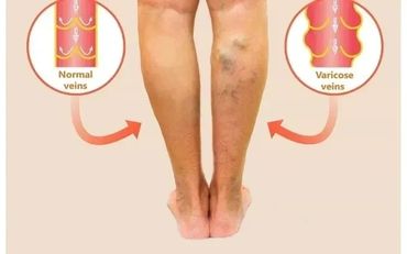
Varicose veins are a condition that occurs when the veins in the legs become enlarged, dilated, and overfilled with blood. Common varicose veins symptoms include swollen and raised veins that often display a bluish-purple color. For those seeking relief, there are various minimally invasive treatment options available to address this condition.
Normal venous flow dynamics and what happens in varicose veins ?
In most cases, varicose veins, which can cause various symptoms, appear on the lower legs. That's because standing and walking upright increases the pressure in the veins of the leg. To push blood back to your heart, veins rely mainly on surrounding muscles and a network of one-way valves. As blood flows through a vein, the cup-like valves alternately open to allow blood through, then close to prevent backflow. For those looking for relief, minimally invasive treatment options are available to address the discomfort associated with varicose veins.
Varicose veins are a condition that occurs when the veins in the legs become enlarged, dilated, and overfilled with blood. Common varicose veins symptoms include swollen and raised veins that often display a bluish-purple color. For those seeking relief, there are various minimally invasive treatment options available to address this condition.
Potential factors leading to varicose veins:
In most cases, varicose veins, which can cause various symptoms, appear on the lower legs. That's because standing and walking upright increases the pressure in the veins of the leg. To push blood back to your heart, veins rely mainly on surrounding muscles and a network of one-way valves. As blood flows through a vein, the cup-like valves alternately open to allow blood through, then close to prevent backflow. For those looking for relief, minimally invasive treatment options are available to address the discomfort associated with varicose veins.
Symptoms of varicose veins:
Bulging veins are a common symptom of varicose veins, presenting as twisted, swollen, rope-like formations that are often blue or purple in color. These veins typically appear just below the surface of the skin on the legs, ankles, and feet. Individuals may also experience heavy, aching, or tender legs, where the muscles feel tired, heavy, or sluggish, especially after prolonged standing. Elevating the legs can provide partial relief. Additionally, itching may occur around the varicose veins, along with swelling in the leg and ankle areas. If left untreated, varicose veins can lead to skin discolorations, presenting as brown or bluish patches on the skin. Severe cases may result in venous ulcers (sores) on the skin, primarily occurring in the lower leg and around the ankle. Occasionally, veins very close to the skin may burst, causing minor bleeding that requires medical attention. Moreover, varicose veins can clot, leading to a condition known as phlebitis, which is characterized by painful, tender, hard, and discolored areas. For those seeking relief, there are minimally invasive treatment options available to address these varicose veins symptoms effectively.
5 Stages of venous disease
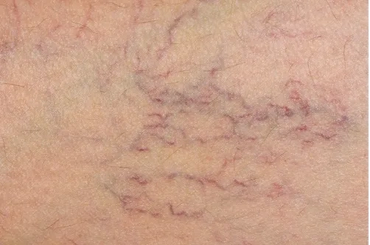
Stage 1: Spider veins or reticular veins appear as tiny threads resembling the veins in a spider web or a cluster-like pattern. These veins are usually asymptomatic and are often linked to a family history of varicose veins, menopause, and hormonal imbalance. While they may not cause discomfort, many seek minimally invasive treatment options to address any potential varicose veins symptoms that may arise.
Varicose veins are a condition that occurs when the veins in the legs become enlarged, dilated, and overfilled with blood. Common varicose veins symptoms include swollen and raised veins that often display a bluish-purple color. For those seeking relief, there are various minimally invasive treatment options available to address this condition.
Diagnosis of varicose veins
In most cases, varicose veins, which can cause various symptoms, appear on the lower legs. That's because standing and walking upright increases the pressure in the veins of the leg. To push blood back to your heart, veins rely mainly on surrounding muscles and a network of one-way valves. As blood flows through a vein, the cup-like valves alternately open to allow blood through, then close to prevent backflow. For those looking for relief, minimally invasive treatment options are available to address the discomfort associated with varicose veins.
Minimally InvasiveTreatment Solutions for Varicose Veins
Specific treatment is determined by the doctor based on
Varicose veins are a condition that occurs when the veins in the legs become enlarged, dilated, and overfilled with blood. Common varicose veins symptoms include swollen and raised veins that often display a bluish-purple color. For those seeking relief, there are various minimally invasive treatment options available to address this condition.
Treatment may include
In most cases, varicose veins, which can cause various symptoms, appear on the lower legs. That's because standing and walking upright increases the pressure in the veins of the leg. To push blood back to your heart, veins rely mainly on surrounding muscles and a network of one-way valves. As blood flows through a vein, the cup-like valves alternately open to allow blood through, then close to prevent backflow. For those looking for relief, minimally invasive treatment options are available to address the discomfort associated with varicose veins.
Varicose veins are a condition that occurs when the veins in the legs become enlarged, dilated, and overfilled with blood. Common varicose veins symptoms include swollen and raised veins that often display a bluish-purple color. For those seeking relief, there are various minimally invasive treatment options available to address this condition.

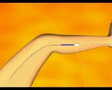
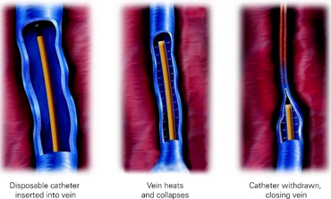
- VenaSeal ( Glue ) Treatment : Venaseal is an advanced minimally invasive treatment using image-guided techniques to ensure precise and effective result for patients suffering from varicose veins uses a specially medical adhesive ( Glue ) to close the diseased vein.

Before and After Treatment
How does the leg drain blood out after treatment of varicose veins?
Varicose veins are a condition that occurs when the veins in the legs become enlarged, dilated, and overfilled with blood. Common varicose veins symptoms include swollen and raised veins that often display a bluish-purple color. For those seeking relief, there are various minimally invasive treatment options available to address this condition.
Post procedure course
In most cases, varicose veins, which can cause various symptoms, appear on the lower legs. That's because standing and walking upright increases the pressure in the veins of the leg. To push blood back to your heart, veins rely mainly on surrounding muscles and a network of one-way valves. As blood flows through a vein, the cup-like valves alternately open to allow blood through, then close to prevent backflow. For those looking for relief, minimally invasive treatment options are available to address the discomfort associated with varicose veins.
Varicose Veins Specialist in Rajkot
Bulging veins are a common symptom of varicose veins, presenting as twisted, swollen, rope-like formations that are often blue or purple in color. These veins typically appear just below the surface of the skin on the legs, ankles, and feet. Individuals may also experience heavy, aching, or tender legs, where the muscles feel tired, heavy, or sluggish, especially after prolonged standing. Elevating the legs can provide partial relief. Additionally, itching may occur around the varicose veins, along with swelling in the leg and ankle areas. If left untreated, varicose veins can lead to skin discolorations, presenting as brown or bluish patches on the skin. Severe cases may result in venous ulcers (sores) on the skin, primarily occurring in the lower leg and around the ankle. Occasionally, veins very close to the skin may burst, causing minor bleeding that requires medical attention. Moreover, varicose veins can clot, leading to a condition known as phlebitis, which is characterized by painful, tender, hard, and discolored areas. For those seeking relief, there are minimally invasive treatment options available to address these varicose veins symptoms effectively.
Varicose Veins Treatment in Rajkot
If you are experiencing varicose veins symptoms, you may have searched for effective varicose veins treatment methods. This video highlights the differences and similarities between minimally invasive treatment options like Endovenous Laser and Glue treatment.
This website uses cookies.
We use cookies to analyze website traffic and optimize your website experience. By accepting our use of cookies, your data will be aggregated with all other user data.